Poikilocytosis is a term used to describe variation in red blood cell (RBC) shape.
Normal, mature RBCs are round and with a zone of central pallor—shown in their deeper pink or red edge and paler centre. It is not uncommon to see a few abnormally-shaped RBCs amongst hundreds and hundreds of cells. Guided by lab protocol, technologists decide whether the abnormality is prevalent enough to be graded and reported.
When a patient has a high quantity of abnormally-shaped cells, there may be a more serious underlying cause. Different shapes are suggestive of different conditions and pathologic processes—I’ve summarized a few of them below.
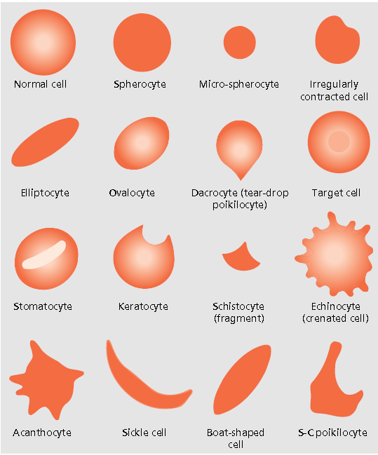
Echinocytes—Also known as burr cells, these RBCs have short and regularly-spaced projections. They are associated with uremia—high levels of urea in the blood due to inadequate kidney function—and pyruvate kinase deficiency. As with all abnormally-shaped red cells, a technologist must consider their frequency both within and across microscopic fields of a peripheral blood film—if a slide is dried too slowly, normal red cells may crenate, giving the appearance of echinocytes and disease when, in actuality, it is just drying artifact.
Elliptocytes—Appear oval or elongated. May be seen in patients with iron deficiency anemia (IDA) and thalassemia major, among other conditions.
IDA develops when there is insufficient iron to meet the body’s needs. Women of childbearing age and children are the most at risk of developing IDA—the former due to monthly menstrual bleeding (increased blood loss leads to an increased need for iron, often not met by an unchanged diet), and the latter linked to insufficient intake during this period of rapid growth.
Be the first to receive the latest BCIT News. Become a subscriber.
Sickle cells—Sickle cell anemia is a hereditary (inherited) condition associated with hemoglobin S, an abnormal form of hemoglobin that causes RBCs to become stiff, and hence difficult to move through blood vessels. The increased blood viscosity and reduced oxygen-carrying capacity of hemoglobin S can lead to hypoxia and recurrent pain episodes.
Target cells—Have hemoglobin concentrated in the centre and around the periphery of red cells, giving the appearance of a target or bullseye. A large number may be seen in patients with liver disease and thalassemia.

Though abnormally-shaped RBCs can be associated with specific disorders, their presence or absence is never diagnostic on its own—many shapes can also result from artifact, such as slow drying of the blood film or a poor-quality sample. At the tech level, one uses their best judgement regarding the significance of what is seen on a slide. Results are correlated before doctors make decisions on clinical diagnosis and monitoring.
Interested in the Medical Laboratory Science full-time diploma program? Join the next online info session at BCIT.